 |
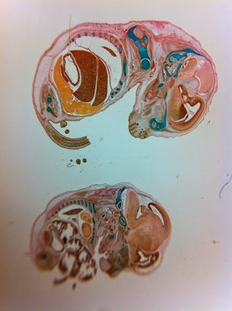
| Histological slides of mouse fetuses in the collection at CELLCentral, UWA. |
Henry is a cat at the house where I am staying, and he waits for me downstairs in the kitchen every morning. He's an empathetic, intelligent cat and even though I don't like cats, I like Henry. I like Henry even more now because he has assisted my research and may be the key to my lab success, in the end. Henry called to everyone in the house last week, late one evening, with some god-awful yowling and once he had everyone's attention, proudly displayed the rat he'd just caught. That very rat accompanied me to the lab the next morning, tightly bound in a plastic container. I sliced it open in the lab and removed its femur bones, which I then extracted the marrow from. It just so happens that I was given a protocol (lab recipe) for isolating osteoblasts from rat bone marrow. I didn't have to use any poor lab rats - this one was a gift from a cat on a mission. This protocol was extremely complex, and I was in the lab alone all day, since Ionat had called in sick. There was nobody around to help me, and so I took a big breath and did the entire thing on my own. I've never cut open a rat before. I was terrified that I'd get in trouble for bringing in a dirty old rat from the outdoors, so I worked quickly to get the bones out and then wrap up the evidence in a bag destined for the biological waste bin. I mastered the art of dressing and removing scalpel blades, since I had to use several on the rat. I asked around for the right kind of pitre dishes and some syringes with different sized needles. I asked about the chemicals listed in the protocol so that I'd have some vague idea of what I was doing. Then I prepared the fresh bones by washing them with ethanol and PBS, then with only two hands (mine), I somehow managed to balance the bones with super fine sterile tweezers and clip the ends off both ends of the bone, attach the needles to the syringes and fill them with PBS, which I then shot through the bones to flush the marrow out into tubes. It was a tiny bit. Then I took my tubes upstairs to the centrifuge, spun them at 1800x/minute and then went back downstairs and pipetted out the fluid until I was down to the tiny bit of marrow at the bottom of the tube. Then I prepared dishes with the right chemicals and plated all the marrow cells and stowed them away in the incubator for the weekend. I felt truly accomplished after all of this. This lady has lab
skillz.
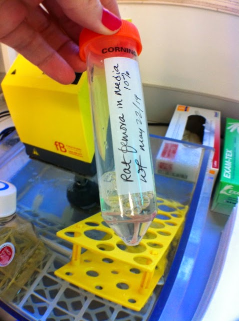 |
| Little rat femurs. |
My microscopy skills have also been improving slowly, as I begin to develop a cell vocabulary. It's really difficult to look for life in a pitre dish when you don't know what you're looking for. So, I can identify red blood cells very easily now, as well as fat cells, air bubbles and crystals. I still have trouble sometimes with seeing my C2C12s, and still have yet to experience a living osteoblast or any other bone cell. I can also identify dead cells, dividing cells and non-cell detritus in the dish. These might seem like simple things but they
really aren't. I've had many false alarms because I got exciting by something, only to discover it was just... an air bubble. Or a dying cell. There were some possible living cells in my cultures at the end of the week last week, confirmed as possibly something by Stuart.
I sent the microscopic digital images I captured on Thursday to an artist collaborator of mine in Montreal, Paloma Dawkins, who is now in the process of drawing and animating them. I'm really excited to see what she'll produce from the images!! Part of the challenge when working within the realm of science and biotechnology is to not just get totally seduced by the technology and lose part of the creativity of the work. So, these little animations are one way to aestheticize the research in a creative manner. Paloma and I worked together last year with great results, so I have every confidence in her. Here are some of the images she's working with and some explanations for what's going on:
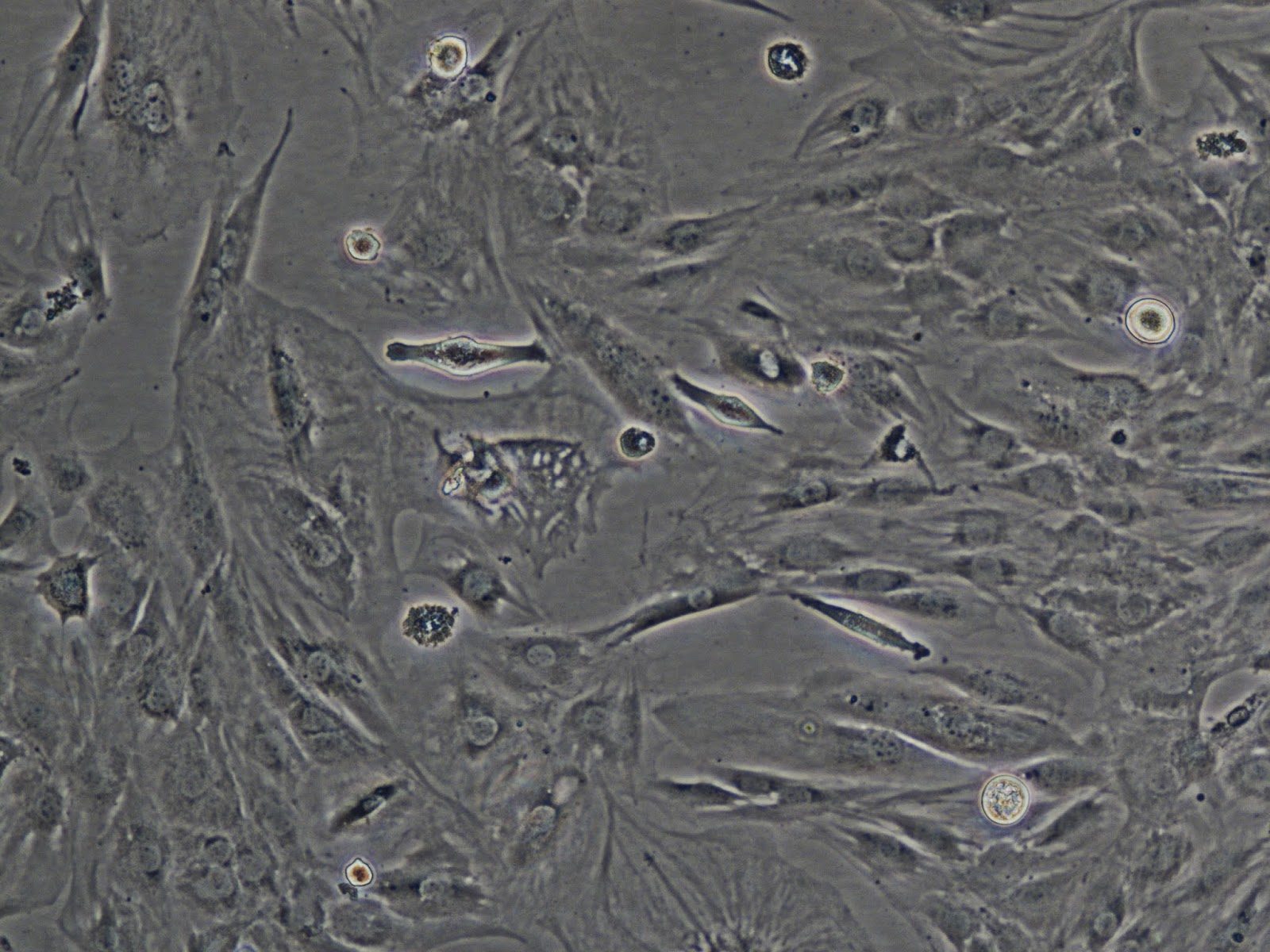 |
| C2C12 (Mighty Mouse) cells. |
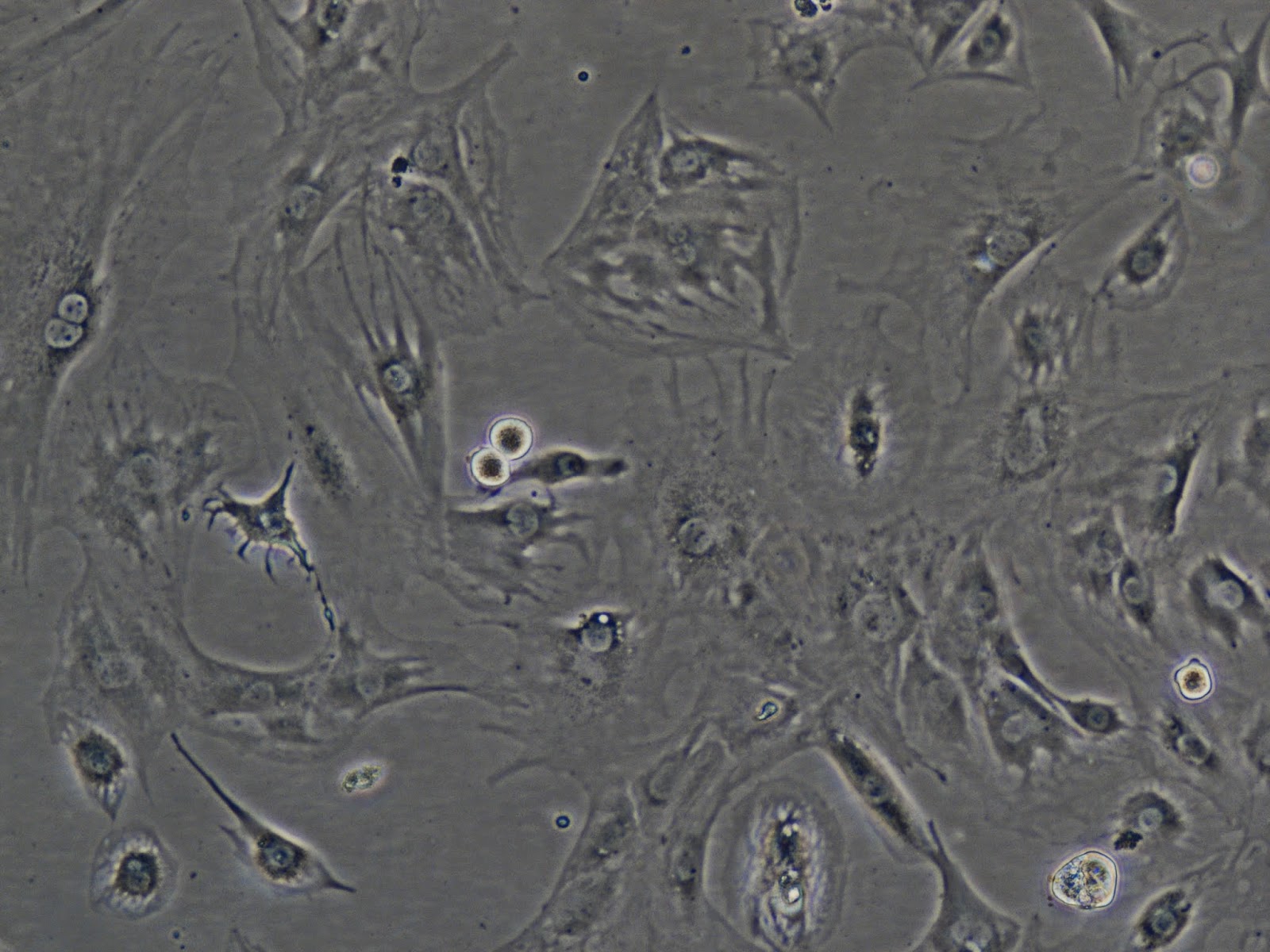 |
| More C2C12s, same dish, different location. |
Aren't they beautiful? This is how they looked last week, but this week they're totally different because they have started to differentiate. The cells with the very bright auras are either about to divide or have just divided (in the case of two next to each other, which you can see in the bottom image). Cells that aren't in the process of division are stuck down to the dish and flattened out, trying to network with each other and their auras aren't as bright. You can see them reaching out, stretching long tendrils towards each other. The circles in the centre of the cells are their nuclei.
THESE are the beautiful universes of bone marrow (from a cow femur):
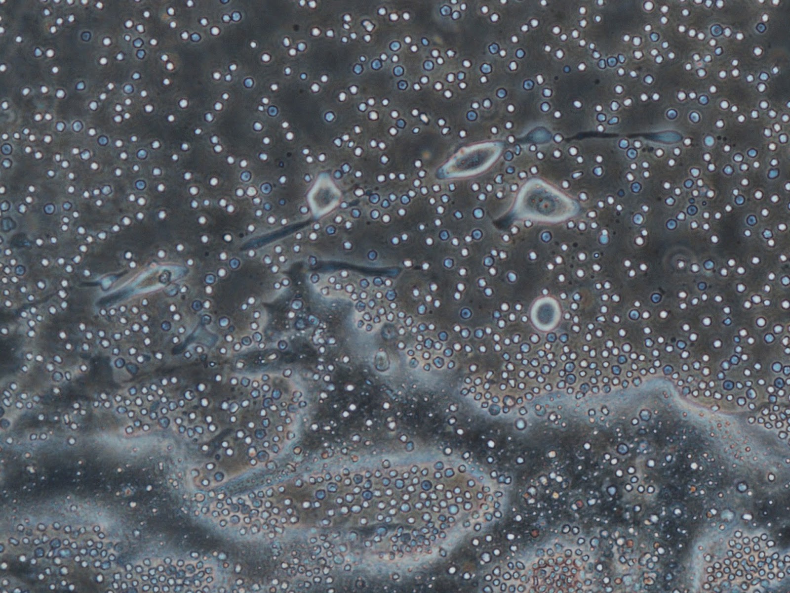 |
| All of the tiny white and blue dots are red blood cells. The white ones are alive, the blue ones are asphyxiating and the ones with black dots in the centre are fully dying. Red blood cells have a short life span and no nucleus. The large cloudy stuff is fat, or oil from burst fat cells. The larger things with auras are what are possibly viable bone cells. Keep your fingers crossed for me. Stuart will look at them this week and tell me what they really are. |
These are the images that Paloma is animating. In this bottom image, you can see nuclei in the cells, which is a good sign (the dark pepper specks inside the cell). This week I'll check the cultures again to see what's still alive. Guaranteed there will be a lot less red blood cells. Will there still be anything else alive? We shall see.
One of my cultures today was full of beautiful growing crystals! This is amazing again because I was growing crystals on hog gut last semester as part of a biomimetic project. All of my lab work is validating everything that I had been working on intuitively before, in my studio/lab. I hope the crystals last long enough for me to photograph them.
Lastly, I began the process of decellularization last week as well, on hog gut. I cut open the intestine, stretched it out into sheets that I pinned down to a styrofoam base and added an enzyme called Trypsin to dissolve all of the cells. It was beautiful. Trypsin is pink, so it coloured the gut until all of the cells were washed away. It's completely white and transparent now. More pics of that later.


No comments:
Post a Comment